Module 02 · Foundations心理学基础
The Brain大脑
By Diane Beck and Evelina Tapia
由 Diane Beck 和 Evelina Tapia
University of Illinois at Urbana-Champaign, University of Illinois
伊利诺伊大学香槟分校,伊利诺伊大学
The human brain is responsible for all behaviors, thoughts, and experiences described in this textbook. This module provides an introductory overview of the brain, including some basic neuroanatomy, and brief descriptions of the neuroscience methods used to study it.
人脑负责本教科书中描述的所有行为、思维和经验。该模块提供了一个简要的大脑概述,包括一些基本的神经解剖学和用于研究大脑的神经科学方法的简要描述。
Learning Objectives 学习目标
- Name and describe the basic function of the brain stem, cerebellum, and cerebral hemispheres.
命名并描述了脑干、小脑和大脑半球的基本功能。 - Name and describe the basic function of the four cerebral lobes: occipital, temporal, parietal, and frontal cortex.
命名并描述了四个大脑叶的基本功能:枕叶、颞叶、顶叶和额叶皮质。 - Describe a split-brain patient and at least two important aspects of brain function that these patients reveal.
描述一个分脑患者及这些患者揭示的至少两个重要的大脑功能方面。 - Distinguish between gray and white matter of the cerebral hemispheres.
区分大脑半球的灰质和白质。 - Name and describe the most common approaches to studying the human brain.
列举并描述研究人类大脑的最常见方法。 - Distinguish among four neuroimaging methods: PET, fMRI, EEG, and DOI.
分辨四种神经影像学方法:PET、fMRI、EEG 和 DOI。 - Describe the difference between spatial and temporal resolution with regard to brain function.
描述脑功能方面的空间分辨率和时间分辨率的区别。
Introduction 介绍
Any textbook on psychology would be incomplete without reference to the brain. Every behavior, thought, or experience described in the other modules must be implemented in the brain. A detailed understanding of the human brain can help us make sense of human experience and behavior. For example, one well-established fact about human cognition is that it is limited. We cannot do two complex tasks at once: We cannot read and carry on a conversation at the same time, text and drive, or surf the Internet while listening to a lecture, at least not successfully or safely. We cannot even pat our head and rub our stomach at the same time (with exceptions, see “A Brain Divided”). Why is this? Many people have suggested that such limitations reflect the fact that the behaviors draw on the same resource; if one behavior uses up most of the resource there is not enough resource left for the other. But what might this limited resource be in the brain?
心理学教科书如果不涉及大脑,就会显得不完整。在其他模块描述的每种行为、思维或体验都必须在大脑中得到实现。对人类大脑的详细了解可以帮助我们理解人类的经验和行为。例如,关于人类认知的一个确立事实是它是有限的。我们不能同时进行两个复杂的任务:不能边读边交谈,边发短信边开车,或者边浏览互联网边听讲座,至少不能成功或安全地这样做。我们甚至不能同时用一只手拍头和一只手摩腹(有例外情况,请参见“大脑分裂”)。为什么会这样呢?许多人认为,这样的限制反映了这些行为利用的是同一资源;如果一个行为耗尽了大部分资源,那么剩下的资源就不足够支持另一个行为。但大脑中可能是什么有限资源呢?
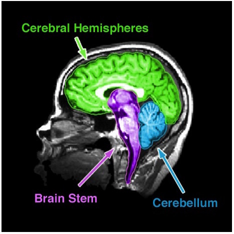
Figure 1. An MRI of the human brain delineating three major structures: the cerebral hemispheres, brain stem, and cerebellum.
图 1. 描绘了人类大脑的磁共振成像,标出了三个主要结构:大脑半球、脑干和小脑。
The brain uses oxygen and glucose, delivered via the blood. The brain is a large consumer of these metabolites, using 20% of the oxygen and calories we consume despite being only 2% of our total weight. However, as long as we are not oxygen-deprived or malnourished, we have more than enough oxygen and glucose to fuel the brain. Thus, insufficient “brain fuel” cannot explain our limited capacity. Nor is it likely that our limitations reflect too few neurons. The average human brain contains 86 billion neurons. It is also not the case that we use only 10% of our brain, a myth that was likely started to imply we had untapped potential. Modern neuroimaging (see “Studying the Human Brain”) has shown that we use all parts of brain, just at different times, and certainly more than 10% at any one time.
大脑利用通过血液输送的氧气和葡萄糖。大脑对这些代谢物的消耗很大,尽管仅占我们总体重量的 2%,但它使用了我们消耗的氧气和热量的 20%。然而,只要我们没有缺氧或营养不良,我们的大脑就有足够的氧气和葡萄糖来供给。因此,不足的“脑燃料”不能解释我们的有限能力。我们的限制也不太可能是由于神经元数量过少。人类平均大脑含有 860 亿个神经元。同样不是我们只使用了大脑的 10%,这是一个可能起源于暗示我们有未开发潜力的迷思。现代神经影像学(参见“研究人脑”)表明,我们在不同时刻使用大脑的各个部分,并且肯定在任何一个时刻使用超过 10%的大脑。
If we have an abundance of brain fuel and neurons, how can we explain our limited cognitive abilities? Why can’t we do more at once? The most likely explanation is the way these neurons are wired up. We know, for instance, that many neurons in the visual cortex (the part of the brain responsible for processing visual information) are hooked up in such a way as to inhibit each other (Beck & Kastner, 2009). When one neuron fires, it suppresses the firing of other nearby neurons. If two neurons that are hooked up in an inhibitory way both fire, then neither neuron can fire as vigorously as it would otherwise. This competitive behavior among neurons limits how much visual information the brain can respond to at the same time. Similar kinds of competitive wiring among neurons may underlie many of our limitations. Thus, although talking about limited resources provides an intuitive description of our limited capacity behavior, a detailed understanding of the brain suggests that our limitations more likely reflect the complex way in which neurons talk to each other rather than the depletion of any specific resource.
如果我们拥有大量的脑能源和神经元,为什么我们的认知能力有限呢?为什么我们不能同时进行更多的事情?最可能的解释是这些神经元的连接方式。例如,我们知道大脑视皮层(负责处理视觉信息的脑部区域)中的许多神经元之间存在一种相互抑制的连接方式(Beck&Kastner,2009)。当一个神经元发射时,它会抑制其他附近神经元的发射。如果两个通过抑制连接的神经元同时发射,那么它们都无法像其他情况下那样强烈地发射。神经元之间的这种竞争行为限制了大脑在同时对视觉信息作出反应方面的能力。类似的竞争性连接方式可能解释了我们许多限制。因此,虽然使用有限资源的说法可以直观地描述我们的有限能力行为,但对大脑的详细理解表明,我们的限制更可能反映出神经元之间复杂的交流方式,而不是任何特定资源的消耗。
The Anatomy of the Brain 大脑的解剖结构
There are many ways to subdivide the mammalian brain, resulting in some inconsistent and ambiguous nomenclature over the history of neuroanatomy (Swanson, 2000). For simplicity, we will divide the brain into three basic parts: the brain stem, cerebellum, and cerebral hemispheres (see Figure 1). In Figure 2, however, we depict other prominent groupings (Swanson, 2000) of the six major subdivisions of the brain (Kandal, Schwartz, & Jessell, 2000).
哺乳动物大脑的分类方法有很多,导致了神经解剖学历史上的一些不一致和模糊的命名方法(Swanson, 2000)。为简单起见,我们将大脑分为三个基本部分:脑干、小脑和脑半球(见图 1)。然而,在图 2 中,我们描述了另外一些主要的分组方法(Swanson, 2000)和大脑的六个主要区域(Kandal, Schwartz, & Jessell, 2000)。
Brain Stem 脑干
The brain stem is sometimes referred to as the “trunk” of the brain. It is responsible for many of the neural functions that keep us alive, including regulating our respiration (breathing), heart rate, and digestion. In keeping with its function, if a patient sustains severe damage to the brain stem he or she will require “life support” (i.e., machines are used to keep him or her alive). Because of its vital role in survival, in many countries a person who has lost brain stem function is said to be “brain dead,” although other countries require significant tissue loss in the cortex (of the cerebral hemispheres), which is responsible for our conscious experience, for the same diagnosis. The brain stem includes the medulla, pons, midbrain, and diencephalon (which consists of thalamus and hypothalamus). Collectively, these regions also are involved in our sleep–wake cycle, some sensory and motor function, as well as growth and other hormonal behaviors.
脑干有时被称为大脑的 "躯干"。它负责维持我们生命的许多神经功能,包括调节呼吸、心率和消化。根据其功能,如果患者的脑干受到严重损伤,就需要 "生命支持"(即使用机器维持生命)。由于脑干在生存中的重要作用,在许多国家,失去脑干功能的人被称为 "脑死亡",但其他国家则要求负责我们意识体验的大脑皮质(大脑半球)有明显的组织损失,才能做出同样的诊断。脑干包括延髓、脑桥、中脑和间脑(由丘脑和下丘脑组成)。这些区域共同参与我们的睡眠-觉醒周期、某些感官和运动功能,以及生长和其他荷尔蒙行为。
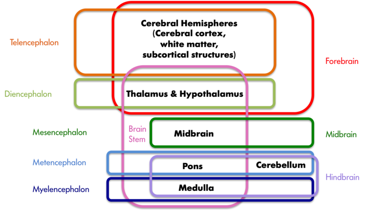
Figure 2. A sample of neuroanatomy nomenclature. The colored boxes indicate the different groupings of the seven structures printed in black, with the labels matching the color of the boxes. The hindbrain, midbrain, and forebrain nomenclature stems from the development of the vertebrate brain; these three areas differentiate early in embryonic development and later give rise to the structures listed in black. These three areas further subdivide into the telencephalon, diencephalon, mesencephalon, metencephalon, and myelencephalon at a later stage of development.
图 2. 神经解剖名称示例。彩色方框表示以黑色字体标出的七个结构的不同分组,标签与方框颜色相匹配。后脑、中脑和前脑的命名来源于脊椎动物大脑的发育;这三个区域在胚胎发育早期即有所区分,并且后来形成了以黑色列出的结构。这三个区域后来在发育的较后阶段进一步分为端脑、间脑、中脑、髓脑和后脑。
Cerebellum 小脑
The cerebellum is the distinctive structure at the back of the brain. The Greek philosopher and scientist Aristotle aptly referred to it as the “small brain” (“parencephalon” in Greek, “cerebellum” in Latin) in order to distinguish it from the “large brain” (“encephalon” in Greek, “cerebrum” in Latin). The cerebellum is critical for coordinated movement and posture. More recently, neuroimaging studies (see “Studying the Human Brain”) have implicated it in a range of cognitive abilities, including language. It is perhaps not surprising that the cerebellum’s influence extends beyond that of movement and posture, given that it contains the greatest number of neurons of any structure in the brain. However, the exact role it plays in these higher functions is still a matter of further study.
小脑是大脑后部的独特结构。希腊哲学家和科学家亚里士多德恰如其分地将其称为“小脑”(希腊语中的“parencephalon”,拉丁语中的“cerebellum”),以区别于“大脑”(希腊语中的“encephalon”,拉丁语中的“cerebrum”)。小脑对协调运动和姿势至关重要。最近的神经影像研究(参见“研究人类大脑”)表明它在一系列认知能力中起着作用,包括语言。也许并不奇怪,小脑的影响超出了运动和姿势的范畴,因为它含有大脑结构中最多的神经元。然而,它在这些更高级功能中的确切作用仍然是需要进一步研究的问题。
Cerebral Hemispheres 脑半球
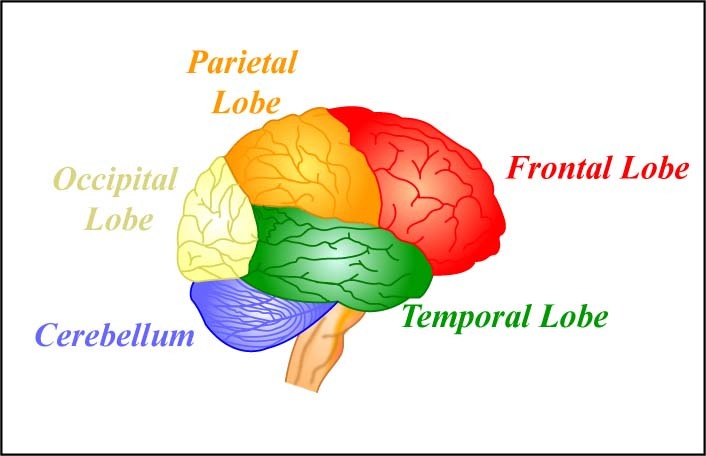
The four lobes of the brain and the cerebellum. [Image: MIT OpenCourseWare, https://goo.gl/RwUEVt, CC BY-NC-SA 2.0, https://goo.gl/Toc0ZF]
脑的四个脑叶和小脑。[图片:MIT 开放式课程网,https://goo.gl/RwUEVt,CC BY-NC-SA 2.0,https://goo.gl/Toc0ZF]
The cerebral hemispheres are responsible for our cognitive abilities and conscious experience. They consist of the **cerebral cortex **and accompanying white matter (“cerebrum” in Latin) as well as the subcortical structures of the basal ganglia, amygdala, and hippocampal formation. The cerebral cortex is the largest and most visible part of the brain, retaining the Latin name (cerebrum) for “large brain” that Aristotle coined. It consists of two hemispheres (literally two half spheres) and gives the brain its characteristic gray and convoluted appearance; the folds and grooves of the cortex are called gyri and sulci (gyrus and sulcus if referring to just one), respectively.
大脑半球负责我们的认知能力和意识体验。它们包括大脑皮质和伴随的白质(在拉丁语中称为“大脑”),以及基底神经节、杏仁核和海马体形成的皮层下结构。大脑皮质是大脑最大、最显著的部分,保留了亚里士多德所创造的“大脑”这个拉丁名字。它由两个半球(字面上的两个半球)组成,赋予大脑其特有的灰色和褶皱的外观;皮质的皱褶和沟槽分别称为脑回和沟(如果只指一个,则为回或沟)。
The two cerebral hemispheres can be further subdivided into four lobes: the occipital, temporal, parietal, and frontal lobes. The occipital lobe is responsible for vision, as is much of the temporal lobe. The temporal lobe is also involved in auditory processing, memory, and multisensory integration (e.g., the convergence of vision and audition). The parietal lobe houses the somatosensory (body sensations) cortex and structures involved in visual attention, as well as multisensory convergence zones. The frontal lobe houses the motor cortex and structures involved in motor planning, language, judgment, and decision-making. Not surprisingly then, the frontal lobe is proportionally larger in humans than in any other animal.
两个大脑半球可以进一步细分为四个叶:枕叶、颞叶、顶叶和额叶。枕叶负责视觉,颞叶的大部分也负责视觉。颞叶还参与听觉处理、记忆和多感官整合(例如,视听的融合)。顶叶承载了体感(身体感受)皮质和参与视觉注意力的结构,还有多感官汇合区域。额叶承载了运动皮质和参与运动规划、语言、判断和决策的结构。因此,相比其他动物,人类的额叶比例较大。
The subcortical structures are so named because they reside beneath the cortex. The basal ganglia are critical to voluntary movement and as such make contact with the cortex, the thalamus, and the brain stem. The amygdala and hippocampal formation are part of the limbic system, which also includes some cortical structures. The limbic system plays an important role in emotion and, in particular, in aversion and gratification.
皮层下结构得名于其位于大脑皮质下方。基底节是自愿运动的关键,因此与皮质、丘脑和脑干相连。杏仁核和海马形成是边缘系统的一部分,该系统还包括一些皮质结构。边缘系统在情绪,特别是厌恶和满足方面起着重要作用。
A Brain Divided 一脑二用
The two cerebral hemispheres are connected by a dense bundle of white matter tracts called the corpus callosum. Some functions are replicated in the two hemispheres. For example, both hemispheres are responsible for sensory and motor function, although the sensory and motor cortices have a contralateral (or opposite-side) representation; that is, the left cerebral hemisphere is responsible for movements and sensations on the right side of the body and the right cerebral hemisphere is responsible for movements and sensations on the left side of the body. Other functions are lateralized; that is, they reside primarily in one hemisphere or the other. For example, for right-handed and the majority of left-handed individuals, the left hemisphere is most responsible for language.
两个大脑半球由一束密集的白质束带连接,称为胼胝体。某些功能在两个半球中复制。例如,两个半球都负责感觉和运动功能,尽管感觉和运动皮层具有对侧(或相反侧)的表征;也就是说,左侧大脑半球负责身体右侧的运动和感觉,右侧大脑半球负责身体左侧的运动和感觉。其他功能是侧化的;也就是说,它们主要存在于一个半球或另一个半球。例如,对于右手和大多数左撇子个体,左半球最负责语言。
There are some people whose two hemispheres are not connected, either because the corpus callosum was surgically severed (callosotomy) or due to a genetic abnormality. These split-brain patients have helped us understand the functioning of the two hemispheres. First, because of the contralateral representation of sensory information, if an object is placed in only the left or only the right visual hemifield, then only the right or left hemisphere, respectively, of the split-brain patient will see it. In essence, it is as though the person has two brains in his or her head, each seeing half the world. Interestingly, because language is very often localized in the left hemisphere, if we show the right hemisphere a picture and ask the patient what she saw, she will say she didn’t see anything (because only the left hemisphere can speak and it didn’t see anything). However, we know that the right hemisphere sees the picture because if the patient is asked to press a button whenever she sees the image, the left hand (which is controlled by the right hemisphere) will respond despite the left hemisphere’s denial that anything was there. There are also some advantages to having disconnected hemispheres. Unlike those with a fully functional corpus callosum, a split-brain patient can simultaneously search for something in his right and left visual fields (Luck, Hillyard, Mangun, & Gazzaniga, 1989) and can do the equivalent of rubbing his stomach and patting his head at the same time (Franz, Eliason, Ivry, & Gazzaniga, 1996). In other words, they exhibit less competition between the hemispheres.
有些人的两个半球没有连接,要么是因为胼胝体被手术切断了(胼胝体切开术)或者因为遗传异常。这些分脑病人帮助我们了解了两个半球的功能。首先,由于感觉信息的对侧表示,如果一个物体只放在左或右视觉半场中,那么分脑病人的右或左半球分别会看到它。实质上,就好像一个人在脑袋里有两个大脑,每个大脑看到世界的一半。有趣的是,由于语言往往定位在左半球,如果我们展示给右半球一个图片并问病人她看到了什么,她会说她什么都没看到(因为只有左半球才能说话,它什么也没看到)。然而,我们知道右半球看到了这个图片,因为如果要求病人在看到图片时按下按钮,由右半球控制的左手会做出反应,尽管左半球否认有任何东西存在。 与完全功能的胼胝体相比,裂脑患者可以同时在右侧和左侧视野中寻找某物(L uck、Hillyard、Mangun 和 Gazzaniga,1989),并且可以同时做等同于抚摸肚子和拍拍头的动作(Franz、Eliason、Ivry 和 Gazzaniga,1996)。换句话说,他们的脑半球之间的竞争较少。
Gray Versus White Matter 灰质与白质
The cerebral hemispheres contain both grey and white matter, so called because they appear grayish and whitish in dissections or in an MRI (magnetic resonance imaging; see, “Studying the Human Brain”). The gray matter is composed of the neuronal cell bodies (see module, “Neurons”). The cell bodies (or soma) contain the genes of the cell and are responsible for metabolism (keeping the cell alive) and synthesizing proteins. In this way, the cell body is the workhorse of the cell. The white matter is composed of the axons of the neurons, and, in particular, axons that are covered with a sheath of **myelin **(fatty support cells that are whitish in color). Axons conduct the electrical signals from the cell and are, therefore, critical to cell communication. People use the expression “use your gray matter” when they want a person to think harder. The “gray matter” in this expression is probably a reference to the cerebral hemispheres more generally; the gray cortical sheet (the convoluted surface of the cortex) being the most visible. However, both the gray matter and white matter are critical to proper functioning of the mind. Losses of either result in deficits in language, memory, reasoning, and other mental functions. See Figure 3 for MRI slices showing both the inner white matter that connects the cell bodies in the gray cortical sheet.
大脑半球包含灰质和白质,因为在解剖或磁共振成像(MRI)中它们呈灰白色。灰质由神经元细胞体组成。细胞体(或细胞体)含有细胞的基因,并负责新陈代谢(维持细胞活力)和合成蛋白质。细胞体是细胞的干活者。白质由神经元的轴突组成,特别是覆盖有髓鞘(脂肪支持细胞)的轴突,其呈白色。轴突传导细胞的电信号,因此对细胞之间的通信至关重要。人们在希望某人更加思考时使用“动动脑筋”的表达方式。这里的“脑筋”可能是指大脑半球,尤其是灰质脑皮层的表面,即凹凸的皮层表面。然而,灰质和白质对大脑正常功能都至关重要。 任何一方的损失都会导致语言、记忆、推理和其他思维功能的缺陷。请参见图 3,其中显示了连接灰色皮层中的细胞体的内部白质的 MRI 切片。
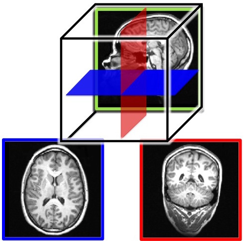
Figure 3. MRI slices of the human brain. Both the outer gray matter and inner white matter are visible in each image. The brain is a three-dimensional (3-D) structure, but an image is two-dimensional (2-D). Here, we show example slices of the three possible 2-D cuts through the brain: a saggital slice (top image), a horizontal slice (bottom left), which is also know as a transverse or axial slice, and a coronal slice (bottom right). The bottom two images are color coded to match the illustration of the relative orientations of the three slices in the top image.
图 3. 人脑的 MRI 切片。每个图像中都可见外部灰质和内部白质。大脑是一个三维结构,但图像是二维的。在这里,我们展示了三种可能的二维脑部切割的示例:矢状切片(顶部图像),水平切片(左下方),也称为横断面切片或轴向切片,以及冠状切片(右下方)。底部两幅图像按照顶部图像中三个切片的相对定位进行了颜色编码以匹配说明图。
Studying the Human Brain 研究人脑
How do we know what the brain does? We have gathered knowledge about the functions of the brain from many different methods. Each method is useful for answering distinct types of questions, but the strongest evidence for a specific role or function of a particular brain area is converging evidence; that is, similar findings reported from multiple studies using different methods.
我们如何了解大脑的功能呢?我们从很多不同的方法中收集关于大脑功能的知识。每种方法都对回答不同类型的问题很有用,但关于特定脑区域的特定角色或功能的最有力证据是收敛证据;也就是说,使用不同方法报告的相似发现。
One of the first organized attempts to study the functions of the brain was phrenology, a popular field of study in the first half of the 19th century. Phrenologists assumed that various features of the brain, such as its uneven surface, are reflected on the skull; therefore, they attempted to correlate bumps and indentations of the skull with specific functions of the brain. For example, they would claim that a very artistic person has ridges on the head that vary in size and location from those of someone who is very good at spatial reasoning. Although the assumption that the skull reflects the underlying brain structure has been proven wrong, phrenology nonetheless significantly impacted current-day neuroscience and its thinking about the functions of the brain. That is, different parts of the brain are devoted to very specific functions that can be identified through scientific inquiry.
大脑功能的最早组织研究之一是骨相学,这是 19 世纪上半叶一门流行的研究领域。骨相学家认为大脑的各种特征,如其不平整的表面,会反映在颅骨上;因此,他们试图将颅骨上的凸起和凹陷与大脑的特定功能相关联。例如,他们会声称一个非常有艺术才华的人的头部凸起和位置与一个非常擅长空间推理的人的不同。虽然颅骨反映大脑内部结构的假设已经被证明是错误的,但骨相学对现代神经科学及其对大脑功能的理解产生了重要的影响。也就是说,大脑的不同部分专门负责非常具体的功能,可以通过科学研究来确认。
Neuroanatomy 神经解剖学
Dissection of the brain, in either animals or cadavers, has been a critical tool of neuroscientists since 340 BC when Aristotle first published his dissections. Since then this method has advanced considerably with the discovery of various staining techniques that can highlight particular cells. Because the brain can be sliced very thinly, examined under the microscope, and particular cells highlighted, this method is especially useful for studying specific groups of neurons or small brain structures; that is, it has a very high spatial resolution. Dissections allow scientists to study changes in the brain that occur due to various diseases or experiences (e.g., exposure to drugs or brain injuries).
大脑的解剖,无论是在动物还是尸体上进行,自公元前 340 年亚里士多德首次发表其解剖以来,一直是神经科学家们的关键工具。此后,随着发现了各种可以突出特定细胞的染色技术,这种方法得以大大改进。由于大脑可以被切成非常薄的片,放在显微镜下观察,并突出显示特定细胞,这种方法特别适用于研究特定群体的神经元或微小的脑结构;换句话说,它具有非常高的空间分辨率。解剖使科学家能够研究由于各种疾病或经历(例如暴露于药物或脑损伤)而发生的大脑变化。
Virtual dissection studies with living humans are also conducted. Here, the brain is imaged using computerized axial tomography (CAT) or MRI scanners; they reveal with very high precision the various structures in the brain and can help detect changes in gray or white matter. These changes in the brain can then be correlated with behavior, such as performance on memory tests, and, therefore, implicate specific brain areas in certain cognitive functions.
也进行了使用活人进行的虚拟解剖研究。在这里,通过使用计算机断层扫描(CAT)或磁共振成像(MRI)扫描仪来对大脑进行成像;它们以非常高的精度显示大脑的各种结构,并且可以帮助检测灰质或白质中的变化。然后可以将大脑中的这些变化与行为(如在记忆测试中的表现)相关联,从而证明特定的脑区在某些认知功能中的重要作用。
Changing the Brain 改变大脑
Some researchers induce lesions or **ablate **(i.e., remove) parts of the brain in animals. If the animal’s behavior changes after the lesion, we can infer that the removed structure is important for that behavior. Lesions of human brains are studied in patient populations only; that is, patients who have lost a brain region due to a stroke or other injury, or who have had surgical removal of a structure to treat a particular disease (e.g., a callosotomy to control epilepsy, as in split-brain patients). From such case studies, we can infer brain function by measuring changes in the behavior of the patients before and after the lesion.
一些研究者会在动物的大脑中引发损伤或切除(即移除)某个部分。如果动物在损伤后的行为发生了变化,我们可以推断被切除的结构对该行为很重要。人类大脑的损伤只能在患者人群中进行研究,也就是那些由于中风或其它伤害而失去了大脑区域,或者因为治疗特定疾病(如癫痫的胼胝体切开术,类似于分脑症患者)而进行了结构切除手术的患者。通过这些病例研究,我们可以通过测量患者在损伤前后行为的变化来推断大脑功能。
Because the brain works by generating electrical signals, it is also possible to change brain function with electrical stimulation. Transcranial magnetic stimulation (TMS) refers to a technique whereby a brief magnetic pulse is applied to the head that temporarily induces a weak electrical current in the brain. Although effects of TMS are sometimes referred to as temporary virtual lesions, it is more appropriate to describe the induced electricity as interference with neurons’ normal communication with each other. TMS allows very precise study of when events in the brain happen so it has a good temporal resolution, but its application is limited only to the surface of the cortex and cannot extend to deep areas of the brain.
因为大脑通过产生电信号来工作,所以通过电刺激也可以改变大脑功能。经颅磁刺激(Transcranial Magnetic Stimulation,简称 TMS)是指在头部施加一种短暂的磁脉冲,从而在大脑中暂时引发弱电流。虽然 TMS 的效果有时被称为暂时的虚拟病变,但更适当的是将引发的电流描述为对神经元之间正常通信的干扰。TMS 允许对大脑事件发生的时间进行非常精确的研究,因此具有较好的时间分辨率,但其应用仅限于皮质表面,无法扩展到大脑的深层区域。
Transcranial direct current stimulation (tDCS) is similar to TMS except that it uses electrical current directly, rather than inducing it with magnetic pulses, by placing small electrodes on the skull. A brain area is stimulated by a low current (equivalent to an AA battery) for a more extended period of time than TMS. When used in combination with cognitive training, tDCS has been shown to improve performance of many cognitive functions such as mathematical ability, memory, attention, and coordination (e.g., Brasil-Neto, 2012; Feng, Bowden, & Kautz, 2013; Kuo & Nitsche, 2012).
经颅直流电刺激(tDCS)类似于 TMS,不同之处在于它直接使用电流而不是通过磁脉冲来诱导电流,通过在颅骨上放置小电极来刺激脑区。低电流(相当于一节 AA 电池)会持续更长时间,比 TMS 更长。当与认知训练结合使用时,tDCS 已被证明可以提高许多认知功能的表现,如数学能力、记忆力、注意力和协调性(例如,Brasil-Neto,2012; Feng,Bowden 和 Kautz,2013; Kuo 和 Nitsche,2012)。
Neuroimaging 神经影像学
Neuroimaging tools are used to study the brain in action; that is, when it is engaged in a specific task. Positron emission tomography (PET) records blood flow in the brain. The PET scanner detects the radioactive substance that is injected into the bloodstream of the participant just before or while he or she is performing some task (e.g., adding numbers). Because active neuron populations require metabolites, more blood and hence more radioactive substance flows into those regions. PET scanners detect the injected radioactive substance in specific brain regions, allowing researchers to infer that those areas were active during the task. Functional magnetic resonance imaging (fMRI) also relies on blood flow in the brain. This method, however, measures the changes in oxygen levels in the blood and does not require any substance to be injected into the participant. Both of these tools have good spatial resolution (although not as precise as dissection studies), but because it takes at least several seconds for the blood to arrive to the active areas of the brain, PET and fMRI have poor temporal resolution; that is, they do not tell us very precisely when the activity occurred.
神经影像工具用于研究大脑在行动中的情况,也就是说,当其从事特定任务时。正电子发射断层扫描(PET)记录大脑中的血液流动。PET 扫描仪可以检测到注射到参与者血液中的放射性物质,参与者在执行某个任务(例如,加法计算)之前或当时注射。因为活跃的神经元群需要代谢物,所以更多的血液,也就是更多的放射性物质流入这些区域。PET 扫描仪可以检测特定脑区中注射的放射性物质,使研究人员能够推断这些区域在任务进行期间是活跃的。功能性磁共振成像(fMRI)也依赖于大脑中的血液流动。然而,该方法测量的是血液中氧气含量的变化,并不需要向参与者注射任何物质。这两种工具都具有良好的空间分辨率(虽然不如解剖学研究精确),但由于血液至少需要几秒钟才能达到大脑的活跃区域,PET 和 fMRI 的时间分辨率较差,也就是说,它们无法精确告诉我们活动发生的时间。
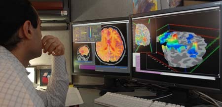
A researcher looking at the areas of activation in the brain of a study participant who had an fMRI scan – areas of brain activation are determined by the amount of blood flow to a certain area – the more blood flow, the higher the activation of that area of the brain. [Image: National Institute of Mental Health, CC0 Public Domain, https://goo.gl/m25gce]
一个研究人员正在观察接受功能性磁共振成像扫描的研究参与者的大脑活跃区域——大脑活跃区域是由特定区域的血流量确定的——血流量越大,该大脑区域的活跃度就越高。【图像:国家心理健康研究所,CC0 公共领域,https://goo.gl/m25gce】
Electroencephalography (EEG), on the other hand, measures the electrical activity of the brain, and therefore, it has a much greater temporal resolution (millisecond precision rather than seconds) than PET or fMRI. Like tDCS, electrodes are placed on the participant’s head when he or she is performing a task. In this case, however, many more electrodes are used, and they measure rather than produce activity. Because the electrical activity picked up at any particular electrode can be coming from anywhere in the brain, EEG has poor spatial resolution; that is, we have only a rough idea of which part of the brain generates the measured activity.
脑电图(EEG)则测量了大脑的电活动,因此它比 PET 或 fMRI 有更高的时间分辨率(以毫秒精度而非秒)。与 tDCS 类似,电极被放置在参与者的头部,当他或她执行任务时。然而,在这种情况下,使用更多电极,并且它们测量而不是产生活动。由于在任何特定电极处捕获的电活动可能来自大脑的任何地方,EEG 的空间分辨率较差;也就是说,我们对大脑的哪个部分产生了测量活动只有一个大致的概念。
Diffuse optical imaging (DOI) can give researchers the best of both worlds: high spatial and temporal resolution, depending on how it is used. Here, one shines infrared light into the brain, and measures the light that comes back out. DOI relies on the fact that the properties of the light change when it passes through oxygenated blood, or when it encounters active neurons. Researchers can then infer from the properties of the collected light what regions in the brain were engaged by the task. When DOI is set up to detect changes in blood oxygen levels, the temporal resolution is low and comparable to PET or fMRI. However, when DOI is set up to directly detect active neurons, it has both high spatial and temporal resolution.
扩散光学成像(DOI)可以给研究人员提供两全其美的好处:高空间和时间分辨率,取决于如何使用。在这里,将红外光照射到大脑中,并测量返回的光。DOI 依赖于当光通过氧合血液时或者遇到活跃的神经元时,光的属性会发生变化的事实。研究人员可以从收集到的光的属性推断哪些脑区被任务激活。当 DOI 被设置为检测血氧水平的变化时,其时间分辨率较低且与 PET 或 fMRI 相当。然而,当 DOI 被设置为直接检测活跃的神经元时,它具有高空间和时间分辨率。
Because the spatial and temporal resolution of each tool varies, strongest evidence for what role a certain brain area serves comes from converging evidence. For example, we are more likely to believe that the hippocampal formation is involved in memory if multiple studies using a variety of tasks and different neuroimaging tools provide evidence for this hypothesis. The brain is a complex system, and only advances in brain research will show whether the brain can ever really understand itself.
因为每个工具的空间和时间分辨率各不相同,所以关于某个特定脑区的功能是什么,最有力的证据来自于多方面的证据交汇。例如,如果多个研究使用不同任务和不同的神经影像工具提供了证据支持,那么我们更有可能相信海马区参与了记忆的过程。大脑是一个复杂的系统,只有脑科研究的进步才能揭示大脑是否能真正了解自己。
Outside Resources 外部资源
Video: Videos of a split-brain patient
视频:分裂大脑患者的视频
Web: Atlas of the Human Brain: interactive demos and brain sections
网络:人脑图谱全集:交互演示和脑部切片
http://www.thehumanbrain.info/
Web: Harvard University Human Brain Atlas: normal and diseased brain scans
网络:哈佛大学人脑图谱:正常和疾病脑部扫描
http://www.med.harvard.edu/aanlib/home.html
Discussion Questions 讨论问题
- In what ways does the segmentation of the brain into the brain stem, cerebellum, and cerebral hemispheres provide a natural division?
大脑如何划分为脑干、小脑和大脑半球会自然地提供一种划分方式? - How has the study of split-brain patients been informative?
对分离脑病患者的研究有何启发? - What is behind the expression “use your gray matter,” and why is it not entirely accurate?
“动动你的脑筋”这个表达背后的含义是什么,为何它并非完全准确? - Why is converging evidence the best kind of evidence in the study of brain function?
为什么收敛证据是研究脑功能的最佳证据? - If you were interested in whether a particular brain area was involved in a specific behavior, what neuroscience methods could you use?
如果您对特定行为是否涉及特定脑区感兴趣,可以使用哪些神经科学方法? - If you were interested in the precise time in which a particular brain process occurred, which neuroscience methods could you use?
如果您对特定脑过程发生的精确时间感兴趣,可以使用哪些神经科学方法?